GCSE / KS4 Science Bundle - Save 10%!
Buy our GCSE/KS4 bundle and save 10%!

Revolutionise the way you learn with Oaka Digital
Designed for Dyslexics, Effective for Everyone
Which type of Oaka product do I need?
How Oaka works in the classroom...
Read what other teachers are saying!
Online learning the Oaka way!
£4.97
In stock
👍 Suitable for AQA, OCR, Edexcel, WJEC, and CCEA exam boards
Buy our GCSE/KS4 bundle and save 10%!

Designed for Dyslexics, Effective for Everyone
✅ Learn or revise complicated concepts easily
✅ Information broken down into short chunks
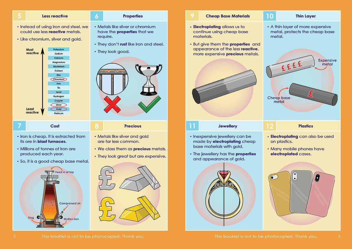
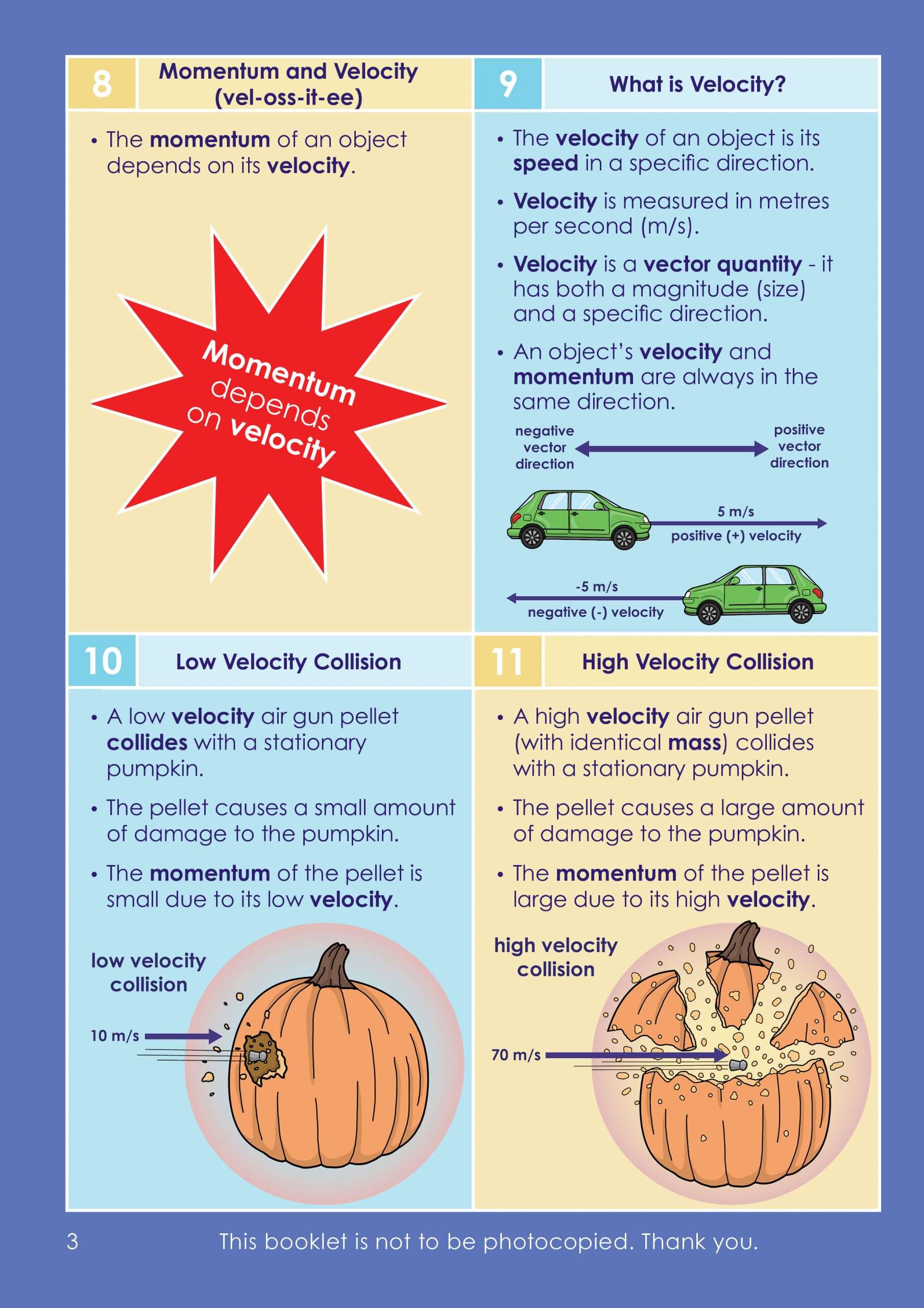
✅ Full-colour illustrations on every page
Using and understanding microscopes is at the heart of many biological experiments. This revision guide, written by Dr Susie Nyman, explains the history, uses and different types of microscopes.
Broken down into bite sized chunks, this Microscopy GCSE revision guide will help students understand (and remember!) the key facts about microscopes and is an essential addition to their knowledge of working scientifically.
Each numbered section is clearly laid out and illustrated to aid understanding, making it more accessible to all students, irrespective of their reading ability, with beautiful images highlighting each key point.
This is a Topic Booklet only – there is no Topic Pack available for this title.
A microscope is a scientific instrument used to magnify objects or organisms beyond the capabilities of the human eye.
The main parts of a microscope include the base, body, nosepiece, objective lenses, stage, light source, focus knobs, and eyepiece.
The history of microscopes dates back to the late 16th century. Dutch spectacle maker Zacharias Janssen is credited with inventing the first compound microscope. Over the years, microscope technology has improved, leading to the creation of electron microscopes, which use electrons instead of light to magnify the specimen.
TEM (Transmission Electron Microscope) and SEM (Scanning Electron Microscope) are types of electron microscopes. TEM uses a beam of electrons transmitted through a thin specimen to produce an image, while SEM uses a beam of electrons that scans the surface of a specimen to produce an image.
TEM provides high-resolution images, with the ability to visualise details down to the atomic level. It is commonly used in materials science, biology, and other fields to study the structure of materials and cells. However, TEM can only be used to study thin specimens that allow electrons to pass through.
SEM, on the other hand, provides images with lower resolution compared to TEM but can be used to study thicker specimens and provides 3D images with excellent surface details. It is commonly used in geology, materials science, and biology to study the surface features of specimens.
Both TEM and SEM are powerful tools for studying the microstructure of materials and organisms, and each has its own advantages and limitations. The choice of which to use depends on the specific research requirements and the type of specimen being studied. Our booklet covers these topics and many more in GCSE microscopes for KS4.

Engaging, full-colour illustrations on every page

Text broken down into bite-sized chunks on a lightly shaded background

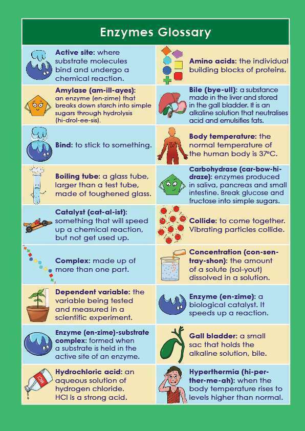
A simple, easy-to-understand glossary of key terms
Topic Booklet
Write Your Own Notes Booklet
Active Learning Q&A Flashcards
Topic Booklet: ✅ x1
Write Your Own Notes Booklet: ✅ x1
Active Learning Q&A Flashcards: ✅ x1
BEST VALUE!
Topic Booklet: ✅ x1
Write Your Own Notes Booklet: ❌
Active Learning Q&A Flashcards: ❌
Topic Booklet: ❌
Write Your Own Notes Booklet: ✅ x1
Active Learning Q&A Flashcards: ❌
Please note, our resources are NOT to be photocopied. Thank you.
Daughter says they are amazing
Absolutely brilliant. Recommending this resource as a revision tool for ALL pupils and spreading the word about Oaka Books resources amongst colleagues in my capacity as dyslexia/dyscalcilia specialist teacher. Can't wait to order and review the ks2 and ks3 publications.
Physics
Biology

In stock